


Volume 3, Issue 2
Pages 1-107 (October 6, 2026)
ISSN: 2584-2153 (Online)
Title: OLCIAS Journal
Intraventricular WHO Grade 1 Glioneuronal Tumor in a Pediatric Patient: A Rare Third Ventricular Presentation
OUADI Hayet1, HARCHAOUI Nabila1, ATROUNE Lynda1 and YOUSFI Yamina2
1: Department of Neurosurgery, Ali Ait Idir Hospital, University of Algiers, Algiers, Algeria
2: Department of Pathology, UHC Nefisa Hammoud, University of Algiers, Algiers, Algeria
*Corresponding Author: OUADI Hayet, Department of Neurosurgery, Ali Ait Idir Hospital, University of Algiers, Algiers, Algeria
Received: August 10, 2026 — Accepted: September 15, 2026 — Published: October 01, 2026
Citation: OUADI Hayet, HARCHAOUI Nabila, ATROUNE Lynda and YOUSFI Yamina. Intraventricular WHO Grade 1 Glioneuronal Tumor in a Pediatric Patient: A Rare Third Ventricular Presentation. OLCIAS Vol.3, Issue 2.
ABSTRACT
Background: Glioneuronal tumors are rare central nervous system (CNS) neoplasms exhibiting both glial and neuronal differentiation. The 2021 WHO classification recognizes multiple entities within this group, most of which are low-grade tumors with a favorable prognosis. Intraventricular locations are particularly uncommon.
Case Presentation: We report the case of a 14-year-old child presenting with intracranial hypertension secondary to a third ventricular mass causing obstructive hydrocephalus. Initial management consisted of ventriculoperitoneal shunting. One year later, the patient developed shunt dysfunction with recurrence of symptoms, requiring external ventricular drainage. Brain MRI confirmed a third ventricular lesion, and the patient subsequently underwent endoscopic subtotal resection combined with endoscopic third ventriculostomy. The postoperative course was favorable, with complete resolution of transient complications, and no adjuvant therapy was administered. Histopathology revealed a low-cellularity glioneuronal tumor composed of oligodendroglial-like cells within a myxoid background, with associated calcifications. Immunohistochemistry showed OLIG2 and GFAP positivity, focal chromogranin A expression, a low Ki-67 index, and negative p53 staining, consistent with a WHO grade 1 glioneuronal tumor.
Conclusion: Intraventricular glioneuronal tumors are rare and diagnostically challenging. Accurate diagnosis relies on the integration of histological and immunohistochemical findings. Endoscopic surgery is a safe and effective approach for deep ventricular locations, with favorable outcomes.
Keywords: glioneuronal tumor; third ventricle; pediatric; hydrocephalus; endoscopic neurosurgery; WHO grade 1
INTRODUCTION
Glioneuronal tumors constitute a distinct group of rare central nervous system neoplasms composed of neoplastic cells with both glial and neuronal differentiation, distinguishing them from purely glial or purely neuronal tumors [ 1]. In the 2021 WHO Classification of Tumors of the Central Nervous System (CNS WHO, 5th edition), glioneuronal tumors are grouped together with neuronal tumors and now encompass a broader spectrum of entities defined by integrated histopathological, immunohistochemical, and molecular features [2].
This updated classification recognizes multiple subtypes of glioneuronal tumors, including ganglioglioma, dysembryoplastic neuroepithelial tumor (DNET), papillary glioneuronal tumor (PGNT), rosette-forming glioneuronal tumor (RGNT), myxoid glioneuronal tumor (MGT), and diffuse glioneuronal tumor with oligodendroglioma-like features and nuclear clusters (DGONC), among others, reflecting a growing understanding of their biology and clinical behavior [2]. Many glioneuronal tumors are considered WHO grade 1 neoplasms owing to their generally indolent course and favorable prognosis, particularly when gross total resection is achievable [ 1].
The intraventricular location of glioneuronal tumors is particularly uncommon; most reported cases involve the lateral or third ventricles and often present with hydrocephalus and symptoms of cerebrospinal fluid obstruction [3]. Magnetic resonance imaging plays a crucial role in preoperative tumor characterization, but definitive diagnosis always relies on histopathological and immunohistochemical analysis demonstrating both glial and neuronal elements [4].
Herein, we report the case of a 14-year-old child with an intraventricular WHO grade 1 glioneuronal tumor, with emphasis on the clinical presentation, imaging findings, surgical approach, and histopathological confirmation, in the context of the current WHO 2021 classification.
CASE PRESENTATION
Clinical presentation and initial management
A 14-year-old child presented with a syndrome of intracranial hypertension characterized by persistent headaches, vomiting, and blurred vision. Emergency brain CT revealed biventricular hydrocephalus associated with an intraventricular lesion occupying the third ventricle, hyperdense on non-contrast images (Figure 1). The patient underwent urgent placement of a ventriculoperitoneal shunt, with effective control of intracranial hypertension (Figure 2).
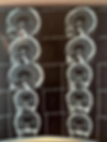
Figure 1. Preoperative brain CT scan in coronal and sagittal sections showing dilation of the lateral ventricles, with the third ventricle occupied by a hyperdense lesion.
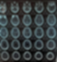
Figure 2. Postoperative brain CT scan in axial sections after placement of a ventriculoperitoneal shunt (VP shunt).
One year after shunt placement, the patient presented again with the same clinical symptoms, secondary to ventriculoperitoneal shunt dysfunction. An external ventricular drain was temporarily placed to relieve intracranial hypertension, and definitive endoscopic management was subsequently decided upon.
Imaging findings
Brain MRI was then performed, confirming an intraventricular lesion within the third ventricle. The lesion was isointense on T1-weighted sequences and predominantly isointense on T2-weighted images, with a peripheral hyperintense rim and a central hypointense component. No contrast enhancement was observed after gadolinium administration (Figure 3).

Figure 3. Brain MRI in axial and sagittal sections, including T1-weighted, non-contrast T1, T2-weighted, and FLAIR sequences.
Surgical management
The patient underwent endoscopic subtotal resection of the intraventricular lesion combined with endoscopic third ventriculostomy.
Intraoperatively, the tumor showed heterogeneous consistency, firm in some areas and friable in others, with a whitish appearance and numerous intratumoral calcifications (Figure 4).

Figure 4. Intraoperative endoscopic images. (A) Lesion visualized through the foramen of Monro. (B) Beginning of debulking. (C) Appearance of the center of the lesion. (D–E–F) Visualization of the floor of the third ventricle after major debulking of the lesion.
Postoperative course
The postoperative course was marked by seizures, which were successfully controlled with antiepileptic medication. The patient also developed transient diabetes insipidus and memory disturbances, both of which resolved during follow-up. Postoperative flow-sensitive brain MRI confirmed patency of the third ventriculostomy (Figure 5).

Figure 5. Brain MRI with flow-sensitive (phase-contrast) sequences, showing partial resection of the lesion and good patency of the ventriculocisternostomy.
Histopathological and immunohistochemical findings
Microscopic examination revealed a moderately cellular glioneuronal proliferation composed of oligodendroglial-like cells arranged in clusters, associated with numerous calcifications. The tumor background was myxoid. A perivascular lymphocytic infiltrate was observed, and fragments of choroid plexus tissue were identified in the specimen.
Immunohistochemical analysis demonstrated:
● Positivity for OLIG2 and GFAP.
● No extravascular CD34 staining.
● Negative NeuN.
● Focal chromogranin A positivity in a small subset of tumor cells.
● Negative p53 expression.
● A low Ki-67 labeling index, indicating low proliferative activity.
Based on the morphological features, immunohistochemical profile, and anatomical location, these findings were consistent with a low-grade glioneuronal tumor, classified as WHO grade 1 according to the 2021 WHO Classification of Tumors of the Central Nervous System. The patient did not receive any adjuvant treatment.
DISCUSSION
Glioneuronal tumors constitute a heterogeneous group of rare central nervous system neoplasms characterized by the coexistence of neoplastic glial and neuronal components, generally associated with low-grade biological behavior and a favorable long-term prognosis [ 1]. The 5th edition of the WHO Classification of Tumors of the Central Nervous System (2021) has substantially reshaped the conceptual framework of these tumors by integrating histological features, immunophenotypic profiles, and molecular alterations into a unified diagnostic approach [ 1,5]. Recent updates have further expanded the spectrum of glioneuronal tumors to include newly recognized entities such as DGONC, MGT, and multinodular and vacuolating neuronal tumor (MVNT), underscoring the increasing heterogeneity of this tumor group and emphasizing that they should no longer be regarded as a single clinicopathological entity but rather as a family of tumors with distinct anatomical predilections, molecular signatures, and clinical behaviors [6].
Clinically, glioneuronal tumors in pediatric patients often present with symptoms of intracranial hypertension, including headaches, vomiting, and visual disturbances, particularly when cerebrospinal fluid pathways are obstructed [7,8]. This clinical pattern was observed in our 14-year-old patient, whose intraventricular tumor caused obstructive hydrocephalus. While seizures are a common presenting symptom in cortically based glioneuronal tumors, intraventricular lesions are more frequently associated with hydrocephalus-related manifestations than with epilepsy [7, 13].
Radiologically, glioneuronal tumors are typically well-circumscribed lesions with variable cystic components and minimal peritumoral edema, reflecting their indolent nature [ 10, 12]. On computed tomography, these tumors may appear isodense or hyperdense and frequently contain intralesional calcifications, a feature clearly identified in our patient and consistently reported in the literature [8, 10, 11]. Magnetic resonance imaging usually shows heterogeneous signal characteristics, with hypointense to isointense areas on T1-weighted sequences and hyperintense regions on T2-weighted and FLAIR images, often corresponding to myxoid or cystic components [ 10– 12]. Contrast enhancement is generally mild or patchy, and diffusion-weighted imaging typically shows no restricted diffusion, supporting the low-grade nature of these tumors [ 12]. However, when arising in intraventricular locations, glioneuronal tumors may closely mimic other ventricular neoplasms such as central neurocytoma, ependymoma, or colloid cyst, making preoperative radiological diagnosis challenging and histopathological confirmation mandatory [8, 13].
Intraventricular glioneuronal tumors are exceptionally rare, with only isolated case reports and small series described in the literature [8, 13]. The third ventricular location observed in our case further broadens the anatomical spectrum of these tumors and underscores the diagnostic difficulty associated with non-cortical presentations [8].
Histopathological examination remains the cornerstone of diagnosis, particularly in atypical intraventricular locations [ 1]. According to the WHO 2021 classification, glioneuronal tumors are defined by the coexistence of neoplastic glial and neuronal components at the microscopic level, distinguishing them from purely glial or purely neuronal tumors [ 1,5]. In our case, microscopic examination revealed a low-cellularity proliferation composed of oligodendroglial-like cells embedded within a prominent myxoid background, associated with abundant calcifications. This histomorphological pattern closely corresponds to that described in myxoid glioneuronal tumors, a recently recognized WHO entity characterized by circumscribed oligodendrocyte-like cells within a myxoid stroma [6, 14].
Immunohistochemistry plays a critical role in confirming the mixed lineage differentiation of glioneuronal tumors. GFAP expression supports glial differentiation, while neuronal markers such as synaptophysin, chromogranin, MAP2, or NeuN may variably label the neuronal component [ 1,6, 14]. In our patient, positivity for OLIG2 and GFAP confirmed glial differentiation, while focal chromogranin positivity supported a neuronal lineage despite negative NeuN staining, a pattern frequently reported in immature or poorly differentiated neuronal components of glioneuronal tumors [6, 14]. The absence of extravascular CD34 staining helped exclude alternative diagnoses such as ganglioglioma, while negative p53 expression and a low Ki-67 proliferation index were consistent with a WHO grade 1 tumor and a favorable prognosis [ 1,7]. Additionally, the perivascular lymphocytic infiltrates observed in our specimen have been described in several low-grade glioneuronal tumors and are thought to reflect their indolent, non-infiltrative biological behavior [7].
Molecular profiling has become an essential adjunct to the modern classification of glioneuronal tumors. Myxoid glioneuronal tumors, in particular, frequently harbor recurrent PDGFRA mutations, which help distinguish them from other low-grade neuroepithelial tumors such as DNET [ 14, 15]. Although comprehensive molecular testing was not available in all reported intraventricular cases, the integrated histopathological and immunophenotypic profile in our patient was sufficient to establish a definitive diagnosis according to WHO 2021 criteria [ 1,5].
From a neurosurgical perspective, surgical resection remains the treatment of choice for glioneuronal tumors. Gross total resection is associated with excellent long-term disease control, although subtotal resection may be acceptable in deep or intraventricular locations given the benign biological behavior of these tumors [ 1, 13]. In our case, an endoscopic approach allowed subtotal tumor resection combined with ventriculocisternostomy, achieving effective tumor debulking and cerebrospinal fluid diversion while minimizing surgical morbidity [9, 16]. Endoscopic approaches to third ventricular tumors have been increasingly adopted in pediatric neurosurgery, offering minimally invasive access, reduced brain retraction, and the possibility of simultaneous CSF pathway restoration compared with transcallosal or transcortical approaches [9, 16]. The risk of recurrence after subtotal resection remains low, and malignant transformation is exceedingly rare, supporting a conservative postoperative strategy with close radiological surveillance [ 1,6].
CONCLUSION
This case illustrates an uncommon intraventricular presentation of a WHO grade 1 glioneuronal tumor in a pediatric patient, expanding the anatomical and clinicopathological spectrum of these rare neoplasms [ 1,8, 13]. It highlights the importance of integrating clinical presentation, advanced imaging, detailed histopathological and immunohistochemical analysis, and a tailored neurosurgical strategy to achieve accurate diagnosis and optimal management [ 1,5,9].
DECLARATIONS
Ethics approval and consent to participate
The study was conducted in accordance with the ethical principles of the Declaration of Helsinki.
Consent for publication
Written informed consent was obtained from the patient's legal guardian for publication of this case report and any accompanying images.
Availability of data and materials
The data supporting the findings of this case report are available from the corresponding author upon reasonable request.
Competing interests
The authors declare that they have no competing interests.
Funding
This research received no specific grant from any funding agency in the public, commercial, or not-for- profit sectors.
Authors' contributions
HO, NH and LA were involved in the surgical management of the patient and drafted the manuscript. YY performed the histopathological and immunohistochemical analysis. NH performed the surgical procedure. All authors read and approved the final manuscript.
Acknowledgements
Not applicable.
REFERENCES
1. Louis DN, Perry A, Wesseling P, et al. The 2021 WHO Classification of Tumors of the Central Nervous System: a summary. Neuro Oncol. 2021;23(8):1231–1251. doi:10.1093/neuonc/noab106.
2. Bale TA, et al. An update on pediatric low-grade gliomas and glioneuronal tumors (WHO 2021 classification). Brain Pathol. 2022.
3. Yano H, Nakayama N, Hirose Y, et al. Intraventricular glioneuronal tumor with disseminated lesions at diagnosis – a case report. Diagn Pathol. 2011;6:119. doi:10.1186/1746-1596-6-119.
4. Park YW, et al. The 2021 WHO Classification of Tumors of the Central Nervous System: imaging and molecular updates. J Magn Reson Imaging. 2023.
5. WHO Classification of Tumours Editorial Board. Central Nervous System Tumours. 5th ed. Lyon: IARC; 2021.
6. Barresi V, Gianno F, Marucci G. Newly recognised tumour types in glioneuronal tumours according to the 5th edition of the CNS WHO Classification. Pathologica. 2022;114:447–454.
7. Blumcke I, Aronica E, Urbach H, et al. A neuropathology-based approach to epilepsy-associated tumors. Acta Neuropathol. 2016;131:1–20.
8. Yano H, et al. Intraventricular glioneuronal tumor: case report. Diagn Pathol. 2011;6:119. doi:10.1186/1746 - 1596-6-119.
9. Rangel-Castilla L, et al. Endoscopic approaches to third ventricular tumors. Neurosurg Focus. 2016;41(3):E6.
10. Koeller KK, Henry JM. Superficial glioneuronal tumors: radiologic-pathologic correlation. Radiographics. 2001;21:1533–1556.
11. Osborn AG. Diagnostic Imaging: Brain. 3rd ed. Elsevier; 2016.
12. Blasel S, et al. Imaging characteristics of glioneuronal tumors. AJNR Am J Neuroradiol. 2010;31:176–184.
13. Hasselblatt M, et al. Intraventricular low-grade neuroepithelial tumors. Acta Neuropathol. 2018;136:273– 285.
14. Chiang JCH, et al. Myxoid glioneuronal tumor: clinicopathologic and molecular characterization. Acta Neuropathol. 2022;144:1–15. doi:10.1007/s00401-022-02492-7.
15. Korshunov A, et al. Molecular genetics of pediatric low-grade glioneuronal tumors. Brain Pathol. 2022.
16. Schroeder HWS. Intraventricular neuroendoscopy. Neurosurg Rev. 2015;38:1–14.