


Volume 3, Issue 2
Pages 1-107 (October 6, 2026)
ISSN: 2584-2153 (Online)
Title: OLCIAS Journal
Post-Traumatic Cervicothoracic Spinal Epidural Hematoma in an 18-Month- Old Infant: A Case Report and Literature Review
HASNAOUI Amina1,*, SIDI MAAMAR Mohamed Amokrane1 and ABDELMALEK Adel1
1: Department of Neurosurgery, Frantz-Fanon University Hospital Center, Blida, Algeria
*Corresponding Author: HASNAOUI Amina, Department of Neurosurgery, Frantz-Fanon University Hospital Center, Blida, Algeria.
Received: August 15, 2026 — Accepted: September 01, 2026 — Published: October 01, 2026
Citation: HASNAOUI Amina, SIDI MAAMAR Mohamed Amokrane and ABDELMALEK Adel. Post-Traumatic Cervicothoracic Spinal Epidural Hematoma in an 18-Month-Old Infant: A Case Report and Literature Review. OLCIAS Vol.3, Issue 2.
ABSTRACT
Post-traumatic spinal epidural hematoma (SEH) is an extremely rare entity in the pediatric population [1]. We report the case of an 18-month-old infant who presented with a cervicothoracic SEH extending from C5 to T4 following a minor domestic fall from a bed. The initial presentation consisted of torticollis and progressive tetraparesis, diagnosed more than one week after the trauma. Spinal magnetic resonance imaging (MRI) confirmed a compressive posterior epidural hematoma. Emergency surgical decompression enabled the evacuation of a dense, highly adherent, and organized epidural clot, leading to complete neurological recovery. This case highlights the importance of considering SEH in young pediatric patients presenting with delayed post-traumatic motor deficits, even in the absence of underlying bone fractures [1].
Keywords: Spinal epidural hematoma, Pediatric neurosurgery, Cervicothoracic spine, Spinal cord compression.
INTRODUCTION
Spinal epidural hematoma (SEH) is a rare neurosurgical emergency [1]. Unlike spontaneous forms—which typically occur in elderly patients or in the presence of predisposing risk factors such as anticoagulant therapy, coagulopathies, or hypertension [1, 2]—its post-traumatic occurrence in infants, particularly without associated osseous lesions, remains exceptional [1, 3]. Early diagnosis in very young children is frequently challenged by subtle or delayed clinical manifestations. We report an original observation managed at the Neurosurgery Department of Frantz Fanon University Hospital in Blida.
CASE PRESENTATION
An 18-month-old male infant with no prior medical history or known bleeding diathesis suffered a domestic trauma (fall from a bed) resulting in a cervicothoracic injury. Following the fall, progressive muscle weakness in all four limbs accompanied by torticollis developed gradually. These symptoms were initially overlooked by the family.
Due to the persistence and progression of the motor deficit over a week after the event, the patient was brought for neurosurgical consultation. Physical examination upon admission revealed a severe cervical spinal syndrome along with tetraparesis.
Emergency spinal MRI demonstrated a compressive, biconvex posterior epidural mass extending from the C5 to T4 vertebral levels. The signal characteristics were consistent with a subacute hematoma, without associated bone or disc injury (Figure 2).
The patient was taken to the operating room urgently following neuroradiological confirmation via a posterior approach. Intraoperatively, a dense, organized, and strongly adherent epidural clot was identified. Clot evacuation was technically delicate but achieved satisfactory spinal cord decompression. No active bleeding source or macroscopic vascular malformation was identified.
The postoperative course was marked by significant clinical improvement, culminating in the complete resolution of tetraparesis and full functional recovery (Figure 3).

Figure 1. Sagittal reconstruction CT scan demonstrating the craniocaudal extent (6.74 cm) and maximum thickness (0.92 cm) of the posterior epidural hematoma compressing the cervicothoracic spinal cord.


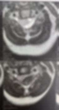
Figure 2. Detailed MRI evaluation reveals a biconvex, posterior epidural collection extending 48 mm in height from C5 to T4, with an average thickness of 13 mm. The signal characteristics (hypointense on T2-weighted images, heterogeneous hyperintense on T1-weighted images, with no enhancement after gadolinium injection) are pathognomonic for an early subacute stage epidural hematoma (intracellular methemoglobin).
Intraoperative photographs are not reproduced in this version.



Figure 3. Comparative sagittal T2-weighted MRI (December 11, 2023 vs. February 20, 2024). This figure illustrates the definitive surgical outcome. The initial scan (December 11) shows severe posterior spinal cord compression by the hematoma (labeled). The follow-up MRI (February 20) demonstrates complete resorption of the cervicothoracic epidural hematoma. The spinal canal diameter is fully restored, showing optimal re-expansion of the spinal cord and restoration of the perimedullary cerebrospinal fluid spaces. No signs of residual compression, spinal cord edema, or suffering are present. The only persisting findings are expected post-surgical fibrous changes in the posterior soft tissues along the approach route.
DISCUSSION
Post-traumatic spinal epidural hematoma accounts for less than 10% of all spinal epidural hematomas according to the classic series by Bruyn and Bosna [1, 4]. Most cases reported in the literature are associated with spinal fractures or fracture-dislocations [1, 3]. The occurrence of a post-traumatic SEH in the absence of bone injury or predisposing bleeding factors is exceptional [1].
Pathophysiology and Delayed Onset
In infants, the high plasticity and ligamentous laxity of the spine allow significant flexion-extension or shearing forces without causing radiologically visible fractures [1]. This shearing mechanism can tear the veins of the posterior epidural plexus [1]. The low pressure within the epidural venous network leads to slow, low-pressure bleeding, gradual clot organization, and progressive meningeal dural stripping [1, 5]. This pathophysiological mechanism accounts for the delayed symptom onset and the diagnostic lag of over one week observed in our patient [1]. Intraoperative findings of a highly organized and condensed clot confirmed the subacute nature of the hemorrhage.
Location and Imaging
According to series by Foo and Rossier, cervical and cervicothoracic locations represent 25% to 43% of all epidural hematomas, developing preferentially within the posterior compartment where the epidural space is anatomically wider [1, 3]. Spinal MRI remains the diagnostic modality of choice [1]. It confirms the epidural location, delineates the craniocaudal extent (C5 to T4 in our patient), and evaluates the severity of spinal cord compression [1]. Hyperintensity on both T1- and T2-weighted images corresponds to extracellular methemoglobin, characteristic of a hematoma older than 7 days [1]. CT scanning may supplement the workup to rule out sagittal fractures not visualized on MRI or plain radiographs [1].
Etiologies to Exclude
In cases of minor trauma, a comprehensive coagulation profile must be systematically performed to rule out an underlying or undiagnosed coagulopathy (e.g., hemophilia) [1, 2]. Occasionally, minor trauma merely unmasks an occult vascular malformation (such as a fistula or angioma), leading several authors to advocate for histopathological examination of the evacuated clot and postoperative spinal angiography [1]. In our patient, coagulation studies were strictly normal.
Prognosis and Pediatric Specificity
Neurological outcome depends on the severity of the preoperative neurological deficit and the promptness of surgical decompression [1, 6]. Foo and Rossier emphasized superior clinical outcomes when surgical decompression is performed early, ideally within the first 36 hours [1, 6]. In the series by Nuti et al., motor recovery reached 95.3% for patients with initial partial deficits, compared to 45.3% when the deficit was complete [1, 7].
However, whereas delayed diagnosis in adults often leads to incomplete functional recovery (as reported by Bencherif et al., where motor recovery remained limited to 3/5 after a 20-day delay) [1], our case demonstrates complete motor recovery in an infant despite a delayed presentation. This highlights the remarkable capacity for neuroplasticity and spinal cord tolerance to subacute compression in infants [1].
CONCLUSION
Post-traumatic spinal epidural hematoma in infants is a rare condition [1]. Diagnosis is frequently delayed owing to progressive clinical presentation and examination difficulties in young pediatric patients [1]. Emergency MRI is the pivotal diagnostic examination [1]. Surgical decompression via hematoma evacuation yields excellent neurological outcomes due to the high recovery potential of the developing pediatric nervous system.
REFERENCES
[1] Bencherif L, Mahfouf L, Abdennebi B. Hématome extradural dorsal post traumatique : à propos d'un cas et revue de la littérature. Journal de Neurochirurgie (Alger). 2008; (7): 20-22.
[2] Bouderka M, Bouaggad A, Ait Benali S, Barrou H, Abassi O. Hématome extradural cervical révélateur d'une hémophilie. Neurochirurgie. 1999; 45(3): 247-249.
[3] Foo D, Rossier AB. Post-traumatic spinal epidural hematoma. Journal of Neurosurgery. 1982; 11: 25-32.
[4] Bruyn GW, Bosma SK. Spinal extradural hematoma. In: Vinken PJ, Bruyn GW, eds. Handbook of Clinical Neurology. Amsterdam: North-Holland; 1976; 26: 1-30.
[5] Clark DB, Bertrand G, Tampieri D. Spontaneous spinal epidural hematoma causing paraplegia: resolution and recovery without surgical decompression. Neurosurgery. 1992; 30(1): 108-111.
[6] Foo D, Rossier AB. Preoperative neurological status in predicting surgical outcome of spinal epidural hematoma. Surgical Neurology. 1981; 15(5): 389-401.
[7] Nuti C, Fotso R, Duthel R, Hatem B, Dumas JP, Brunon J. Revue des hématomes épiduraux non traumatiques du rachis : présentation de 20 cas. Neurochirurgie. 2003; 49(6): 563-570.